Title
題目
PARADISE: Personalized and regional adaptation for HIE disease ?identification and segmentation
PARADISE:用于新生兒缺氧缺血性腦病(HIE)疾病識別與分割的個性化和區域適應性方法
1
文獻速遞介紹
缺氧缺血性腦病(HIE)是一種影響約千分之一到千分之五足月新生兒的腦功能障礙疾病(格雷厄姆等人,2008;李等人,2013)。HIE病變檢測是HIE臨床治療中的關鍵步驟。它有助于更準確地評估預后、更好地理解神經癥狀,并及時預測該人群對治療的反應。人工智能(AI)的興起為在臨床常規獲取的磁共振圖像(MRI)中客觀、準確地發現HIE病變帶來了希望。當前的方法主要集中于大腦腫瘤(門澤等人,2014;馬加扎和維里里,2021;哈瓦埃等人,2017;蒂瓦里等人,2020)、急性中風病變(邁爾等人,2017;克萊里格斯等人,2020;劉等人,2021a)等大的局灶性病變以及其他醫學圖像分割任務(利特延斯等人,2017;雷等人,2020;沙姆沙德等人,2022;卡姆尼察斯等人,2017;周等人,2019b;阿洛姆等人,2019;奧克泰等人,2018;向等人,2020;韓等人,2022;瓦拉納拉蘇和帕特爾,2022;顧等人,2019;張等人,2018;王等人,2020a,2022;曹等人,2021;鮑等人,2021;何等人,2023)。然而,針對HIE的MRI人工智能研究僅有一兩項,且使用的是專有單中心數據,樣本量較小(幾十例),報告的準確率也不理想(墨菲等人,2017)。此外,將現有的醫學成像方法直接應用于HIE并不能得到令人滿意的結果。這主要是因為與HIE病變相比,大多數腦腫瘤和急性中風病變是局灶性的,占據的腦體積要大得多。然而,在MRI中觀察到的與HIE相關的腦部異常(如圖1所示)通常是彌散性(即多灶性)且微小(小于1%)的,超過50%的HIE患者病變影響的腦體積小于1%(鮑等人,2023)。如圖2的第一行和第二行所示,大多數腦腫瘤患者1表現為局灶性病變,其占腦總體積的中位數通常為6.1%。在急性中風病例中2,這一中位數增加到12.5%。形成鮮明對比的是,HIE病變僅占腦總體積中位數的0.6%。大的局灶性病變分割與HIE微小彌散性病變分割在Dice重疊率方面存在明顯的性能差距。如圖2的第三行所示,大的局灶性病變分割性能約為80%,而微小彌散性病變的分割性能約為50%。因此,迫切需要專門針對占HIE病例超過50%的微小彌散性HIE病變分割的算法。設計用于HIE的機器學習方法面臨以下挑戰:(a)病變大小和位置差異大,彌散性病變體積從幾百到幾萬立方毫米不等,腦梗死可發生在腦葉、小腦和腦干等任何腦區(鮑等人,2023)。(b)HIE彌散性病變的多灶性分布和邊界不清晰,這些模糊像素可能會使算法產生混淆。(c)病變體素與正常體素之間的嚴重不平衡會使結果偏向正常類別,因為簡單地假設所有體素都是正常的只會導致一小部分體素被誤分類。算法在損失函數中缺乏足夠的梯度來學習如何聚焦并正確分類病變體素。這導致假陽性(專家標記為正常但算法錯誤分類為病變的體素)比假陰性(專家識別為病變但算法錯誤分類為正常的體素)更多。本文提出個性化區域適應網絡(ParadiseNet),通過精心設計的模塊來應對上述挑戰。ParadiseNet的設計靈感來自臨床診斷流程,在將機器學習算法與HIE檢測相結合的同時,將臨床專家置于設計核心位置。ParadiseNet由三個模塊組成。首先,受醫生診斷流程(包括全局診斷和對局部區域的仔細觀察)啟發,我們設計了全局 - 局部學習模塊,該模塊可以捕捉全局上下文和局部更精細的細節。由于小病變區域在圖像塊上占比更大,局部流被設計為在局部圖像塊上運行。因此,全局 - 局部學習模塊有助于緩解數據不平衡問題。其次,我們設計了漸進不確定性學習模塊,該模塊學習生成分割圖,并從較粗尺度到較細尺度逐步細化不確定的病變區域。第三,我們設計了自進化學習模塊,通過在輸出病變分割圖和輸入之間添加反饋回路來增強ParadiseNet,使單個網絡模型能夠在不增加參數的情況下對病變分割圖進行迭代細化。與現有方法相比,我們為微小彌散性病變分割任務建立了一種新穎且專門的網絡結構。本文的主要貢獻總結如下: - 我們引入了ParadiseNet,旨在應對缺氧缺血性腦病(HIE)中微小彌散性病變分割這一具有挑戰性的任務。 - 我們提出了三個互補組件,每個組件都受臨床診斷指南啟發,以有效應對微小病變分割中病變檢測不準確的挑戰。 ?- 我們證明,與UNet相比,在整個數據集上,ParadiseNet顯著提高了HIE病變分割的Dice準確率,Dice系數提高了3%,歸一化表面距離(NSD)提高了3%。值得注意的是,對于受影響區域小于總體積1%的微小彌散性病變,ParadiseNet的Dice系數提高了4%,NSD提高了6%;對于小于2%的病變區域,Dice系數提高了4% ,NSD提高了5%。這對HIE尤為重要,因為超過一半的病例病變占據的區域小于1%。 本文其余部分的組織如下:第2節回顧醫學圖像分割的相關工作。第3節詳細介紹我們提出的方法ParadiseNet。第4節描述實驗設置,第5節展示定量和可視化結果。最后,第6節對本文進行總結并討論未來的研究方向。
01
文獻速遞介紹
Hypoxic ischemic encephalopathy (HIE) is a brain dysfunction occurring in approximately 1-5/1000 termborn neonates. Accurate segmentation of HIE lesions in brain MRI is crucial for prognosis and diagnosis but ?presents a unique challenge due to the diffuse and small nature of these abnormalities, which resulted in ?a substantial gap between the performance of machine learning-based segmentation methods and clinical ?expert annotations for HIE. To address this challenge, we introduce ParadiseNet, an algorithm specifically ?designed for HIE lesion segmentation. ParadiseNet incorporates global–local learning, progressive uncertainty ?learning, and self-evolution learning modules, all inspired by clinical interpretation of neonatal brain MRIs. ?These modules target issues such as unbalanced data distribution, boundary uncertainty, and imprecise lesion ?detection, respectively. Extensive experiments demonstrate that ParadiseNet significantly enhances small lesion ?detection (< 1%) accuracy in HIE, achieving an over 4% improvement in Dice, 6% improvement in NSD ?compared to U-Net and other general medical image segmentation algorithms.
缺氧缺血性腦病(HIE)是一種腦功能障礙疾病,約每1000名足月新生兒中就有1 - 5名患病。在腦部磁共振成像(MRI)中準確分割HIE病變對于預后和診斷至關重要,但由于這些病變具有彌散性和微小性的特點,給分割帶來了獨特挑戰,導致基于機器學習的分割方法與臨床專家對HIE的標注結果之間存在較大差距 。為應對這一挑戰,我們引入了ParadiseNet算法,這是一種專門為HIE病變分割設計的算法。ParadiseNet融合了全局 - 局部學習、漸進不確定性學習和自進化學習模塊,其設計靈感均源于對新生兒腦部MRI的臨床解讀。這些模塊分別針對數據分布不均衡、邊界不確定性和病變檢測不精確等問題。大量實驗表明,ParadiseNet顯著提高了HIE中微小病變(<1%)的檢測準確率,與U-Net及其他通用醫學圖像分割算法相比,Dice系數提高了4%以上,歸一化表面距離(NSD)提高了6% 。
Conclusion
結論
We address the challenge of detecting small diffuse lesions in HIE, ?a task that has not been well explored. Our proposed ParadiseNet, designed by clinical guidance, which incorporates global–local learning, ?progressive uncertainty learning, and self-evolution learning modules, ?effectively mitigates issues related to unbalanced data distribution, ?boundary uncertainty, and inaccurate lesion detection, as demonstrated ?by comprehensive experimental analysis. ParadiseNet has achieved ?state-of-the-art performance, marking significant advancements over ?existing methods in HIE small diffuse lesion task. Looking ahead, we ?plan to apply and further enhance ParadiseNet for the detection of ?small diffuse lesions in other diseases such as multiple sclerosis and ?subacute ischemic stroke.
我們針對缺氧缺血性腦病(HIE)中微小彌散性病變檢測這一尚未充分探索的任務展開研究。通過臨床指導設計的ParadiseNet融合了全局-局部學習、漸進不確定性學習和自進化學習模塊,經全面實驗分析驗證,該模型有效緩解了數據分布不平衡、邊界不確定性和病變檢測不精確等問題。ParadiseNet已實現了當前最優性能,標志著在HIE微小彌散性病變任務中較現有方法取得了顯著進展。 ? 展望未來,我們計劃將ParadiseNet應用于多發性硬化、亞急性缺血性中風等其他涉及微小彌散性病變的疾病檢測,并進一步優化其性能。具體方向包括: ? 1. 多模態數據融合:結合功能磁共振成像(fMRI)或彌散張量成像(DTI)等多模態信息,提升低對比度病變的特征表征能力。 ? 2. 模型輕量化:通過知識蒸餾或架構優化,在保持精度的同時降低計算復雜度,推動臨床實時部署。 ? 3. 跨中心泛化研究:在更大規模、多中心數據集上驗證模型魯棒性,解決不同成像設備和協議帶來的異質性問題。 ? 4. 臨床決策支持整合:將分割結果與預后預測模型結合,構建從病變檢測到治療方案推薦的全鏈條AI系統,輔助醫生制定個性化診療策略。 ? 通過上述探索,ParadiseNet有望為更多涉及微小彌散性病變的神經疾病提供精準、高效的診斷工具,推動人工智能在精準醫療中的實際應用。
Results
結果
5.1. Comparisons with STOA general algorithms ParadiseNet consistently achieves state-of-the-art performance ?on small diffuse lesion segmentation. In our study, we comparedParadiseNet with widely-utilized deep learning architectures in medical ?imaging. The results are summarized in Table 2, under two distinct settings: the whole dataset, where evaluation metrics are calculated across ?the entire dataset to measure overall quality, and small diffuse lesion ?cases, where metrics are specifically computed for patients with small ?diffuse lesions (lesion percentage less than 1% and 2%). Compared ?to existing methods, our model demonstrates superior performance, ?particularly in small diffuse lesion regions, highlighting its efficacy in ?addressing this challenging detection task. (1) Whole dataset setting: As ?indicated in Table 2, ParadiseNet achieves a mean Dice coefficient of ?0.61, which is a 3% increase over the closest competing methods in the ?whole dataset for lesion region prediction accuracy. Moreover, it shows ?a notable improvement in NSD, with a mean of 0.72, corresponding ?to a 2% enhancement in boundary prediction accuracy compared to ?other UNet variants. The sensitivity of ParadiseNet is remarkable, with ?a mean of 0.64, reinforcing its ability to detect true positives effectively. ?Simultaneously, its specificity remains exceptionally high at a mean of ?0.9989, indicating a low rate of false positives in lesion identification. ?(2) On small diffuse lesions: Notably, ParadiseNet exhibits significant ?improvements in small diffuse lesion cases, especially with a Dice ?increase of 5% and NSD of 5% for lesions less than 1% over nnUNet, ?and a Dice and NSD increase of 4% for lesions less than 2% with ?most methods. These results underscore ParadiseNet’s capability in ?managing unbalanced data distribution and its precision in delineating ?lesion boundaries for small diffuse lesions. The predictive performances ?of different methods are also visually presented in Fig. 5. We visualized ?slices from different patients, each displaying various lesioned areas. ?The Dice and NSD metrics for the whole brain volume are displayed ?beneath each image. These visualizations clearly demonstrate that ParadiseNet significantly and consistently outperforms in terms of Dice and ?NSD metrics.
5.1 與最先進通用算法的比較 ? ParadiseNet在微小彌散性病變分割任務中持續實現了最先進的性能。在本研究中,我們將ParadiseNet與醫學影像中廣泛使用的深度學習架構進行了比較。結果總結于表2,分為兩種不同場景:全數據集場景(計算整個數據集的評估指標以衡量整體性能)和微小彌散性病變場景(專門針對病變占比小于1%和2%的患者計算指標)。與現有方法相比,我們的模型表現出更優的性能,尤其在微小彌散性病變區域,突顯了其應對這一挑戰性檢測任務的有效性。 ? #### (1)全數據集場景 ? 如表2所示,ParadiseNet的平均Dice系數達0.61,相比全數據集中最接近的競爭方法,病變區域預測準確率提升了3%。此外,其歸一化表面距離(NSD)顯著改善,平均值為0.72,相比其他UNet變體,邊界預測準確率提升了2%。ParadiseNet的敏感性表現卓越,平均值為0.64,證實了其有效檢測真陽性病變的能力。同時,其特異性維持在極高水平,平均值為0.9989,表明病變識別中的假陽性率極低。 (2)微小彌散性病變場景 ? 值得注意的是,ParadiseNet在微小彌散性病變病例中表現出顯著改進:對于占比小于1%的病變,其Dice系數相比nnUNet提升5%,NSD提升5%;對于小于2%的病變,相比大多數方法,Dice和NSD均提升4%。這些結果凸顯了ParadiseNet在處理數據分布不平衡問題上的能力,以及在勾勒微小彌散性病變邊界時的精確性。 ? 不同方法的預測性能也在圖5中進行了可視化展示。我們選取了不同患者的切片,每例顯示不同的病變區域,圖像下方標注了全腦體積的Dice和NSD指標。這些可視化結果清晰表明,ParadiseNet在Dice和NSD指標上顯著且持續優于其他方法。
Figure
圖
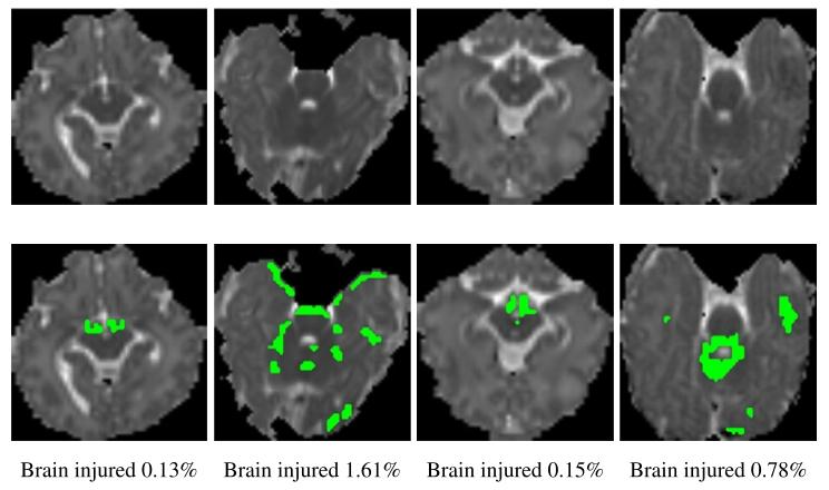
Fig. 1. Hypoxic ischemic encephalopath (HIE) is a typical small diffuse lesion dataset. ?Legion regions in half of the patients in the HIE dataset are less than 1%.
圖1. 缺氧缺血性腦病(HIE)是典型的微小彌散性病變數據集。HIE數據集中半數患者的病變區域占比小于1%。
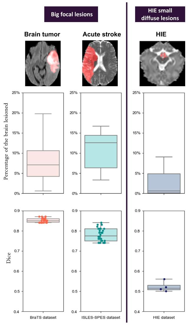
Fig. 2. Big focal lesion detection on ISLES-SPES (Maier et al., 2017; Kistler et al., 2013) ?and BraTS (Menze et al., 2014) vs HIE (Bao et al., 2023) small diffuse detection. The ?systematic gap in the Dice accuracies and methods of big focal lesion segmentation ?versus HIE small diffuse lesion segmentation are displayed. The first row visualizes a ?MRI image of a randomly-chosen patient and the expert-annotated lesions (red regions) ?from each dataset. The second row quantifies the percentage of brain volume lesioned ?across all patients within the dataset. In the third row, each dot represents the mean ?Dice accuracy reported of a method. A clear gap in lesion segmentation accuracies ?and number of methods are illustrated for small diffuse lesions, as noted by the ?gray horizontal stripe in the third row. (See ISLES-SPES and BraTS challenge sites for ?performance reporting in our Figure, and performances on HIE dataset are generated ?by us using deep learning methods designed for other lesion segmentation or from ?literatures).
圖2. ISLES-SPES(Maier等人,2017;Kistler等人,2013)和BraTS(Menze等人,2014)的大灶性病變檢測與HIE(Bao等人,2023)微小彌散性病變檢測的對比。圖中展示了大灶性病變分割與HIE微小彌散性病變分割在Dice準確率和方法上的系統性差距。 ? - 第一行:隨機選取的各數據集患者MRI圖像及專家標注的病變區域(紅色)。大灶性病變(如腦腫瘤、急性中風)呈現明顯局灶性分布,而HIE病變表現為多灶性、微小且彌散的特點。 ? - 第二行:各數據集中所有患者的病變體積占腦體積的百分比。腦腫瘤和急性中風病變體積占比中位數分別為6.1%和12.5%,而HIE病變僅為0.6%,突顯HIE病變的“微小性”。 ? - 第三行:不同方法的平均Dice準確率。大灶性病變分割準確率約80%,而HIE微小彌散性病變僅約50%,灰色橫條標注了兩類任務的顯著性能差距,表明現有方法對微小病變分割效果顯著不足。
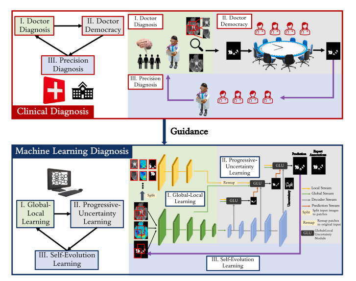
Fig. 3. Overview of our proposed ParadiseNet. ParadiseNet consists of three learning modules: global–local learning module, progressive-uncertainty learning module and selfevolution learning module.It mimics the concepts of doctor diagnosis, doctor democracy, and precision diagnosis in the clinical diagnosis.
圖 3. 我們提出的 ParadiseNet 概述。該網絡包含三個學習模塊:全局 - 局部學習模塊、漸進式不確定性學習模塊和自進化學習模塊,其設計靈感源于臨床診斷中的 “醫生診斷”“多專家會診” 和 “精準診斷” 概念。
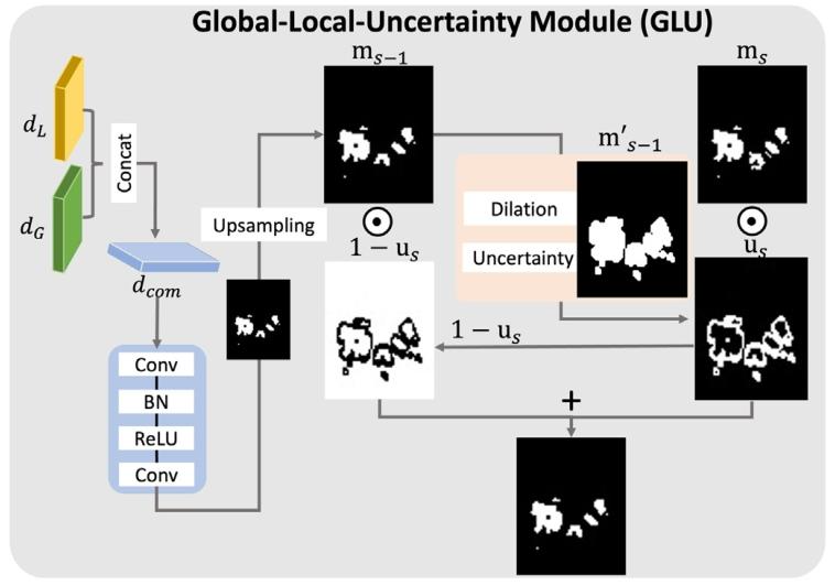
Fig. 4. Global–local-uncertainty learning module.
圖4. 全局-局部-不確定性學習模塊
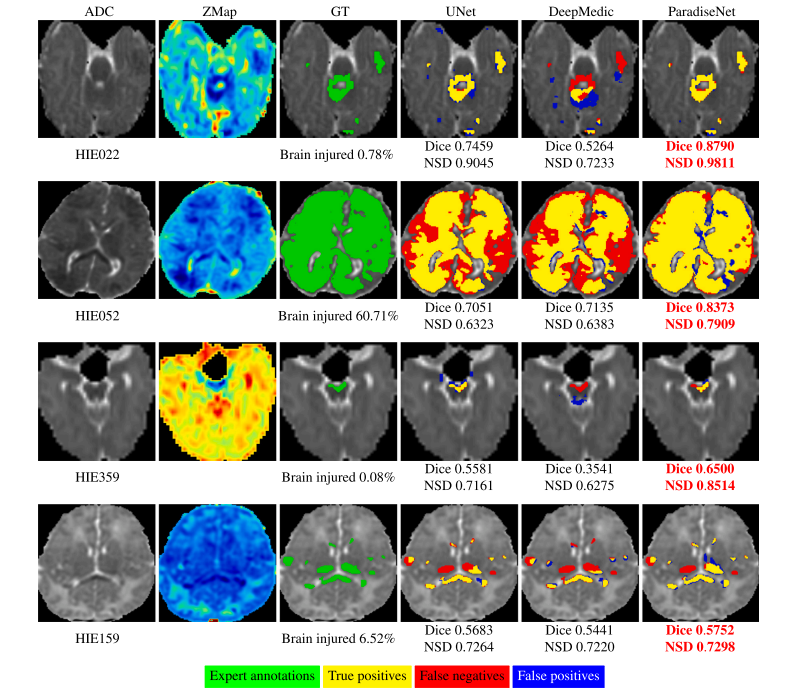
Fig. 5. Lesion segmentation results of different methods. Each row is a patient with different percentages of the brain being lesioned. Compared with existing methods, ParadiseNet ?higher accuracy in lesion regions with different percentage.
圖5. 不同方法的病變分割結果。每行展示一位腦損傷占比不同的患者。與現有方法相比,ParadiseNet在不同損傷比例的病變區域中均表現出更高的準確性。
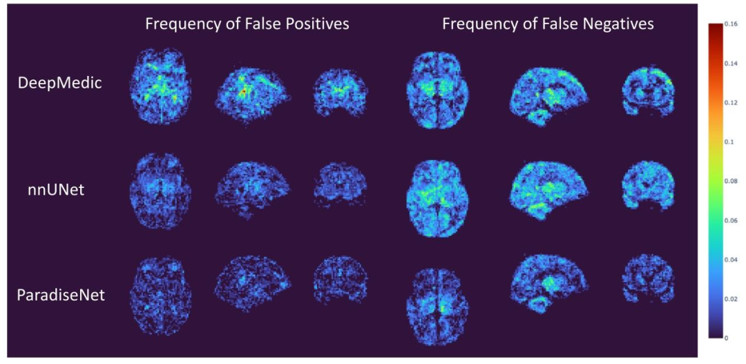
Fig. 6. Frequency maps of false-negative and false-positive detections in 133 patients. The value at each voxel quantified the percentage of patients out of our 133 patients that ?had false-positive (left column) or false-negative (right column) lesion detection using DeepMedic (top row), nnUNet (middle row) and proposed ParadiseNet (bottom row). The ?frequency is color-coded according to the vertical color bar on the right side.
圖 6. 133 例患者假陰性和假陽性檢測的頻率圖。每個體素的值表示 133 例患者中使用 DeepMedic(頂行)、nnUNet(中間行)和 proposed ParadiseNet(底行)檢測到假陽性(左列)或假陰性(右列)病變的患者百分比。頻率根據右側的垂直色條進行顏色編碼。
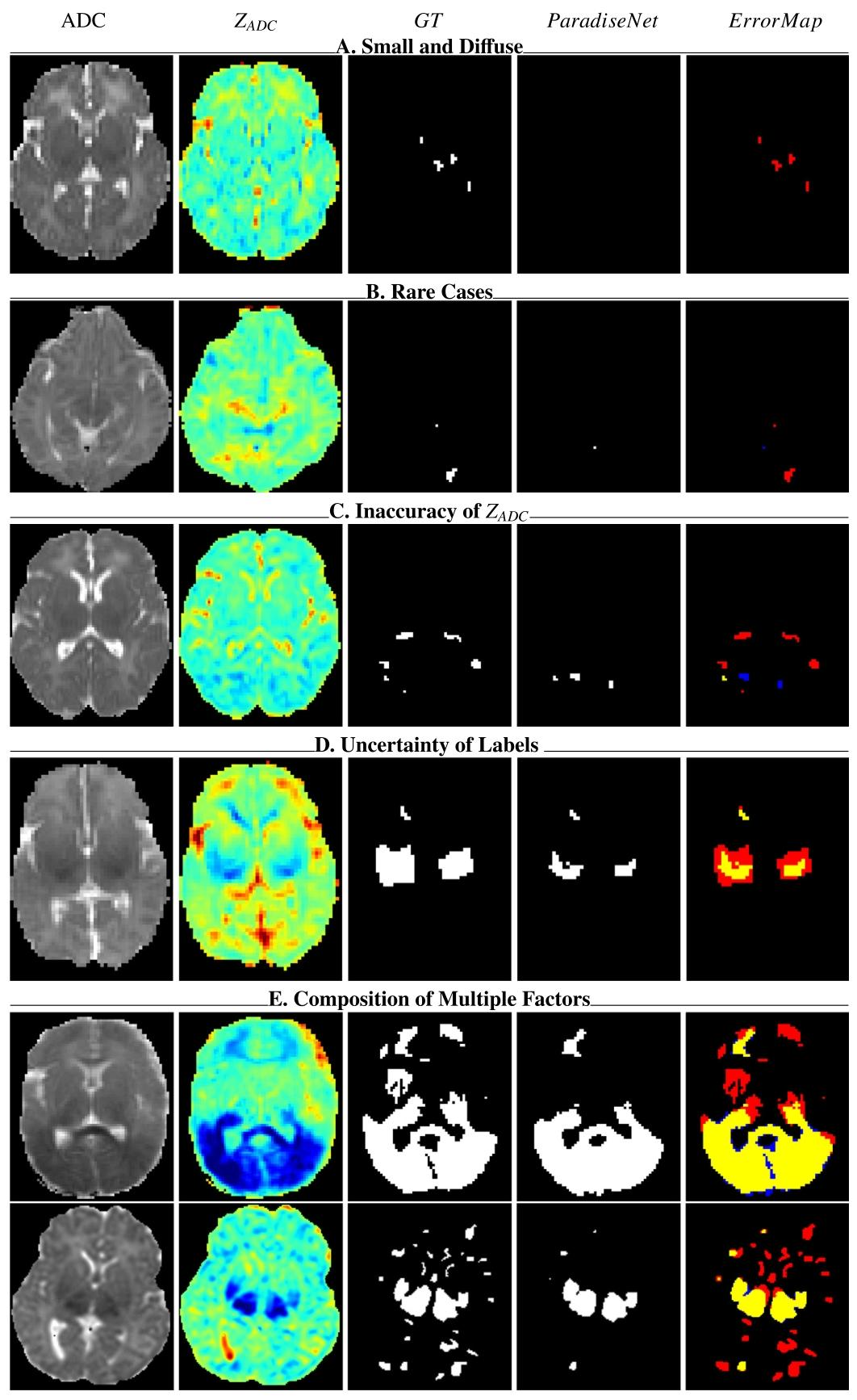
Fig. 7. Failure cases in HIE. Yellow indicates true positives, red denotes false negatives, and blue denotes false positives.
圖 7. HIE 分割失敗案例。黃色表示真陽性(正確檢測的病變),紅色表示假陰性(漏檢的病變),藍色表示假陽性(誤檢的正常區域)。
Table
表
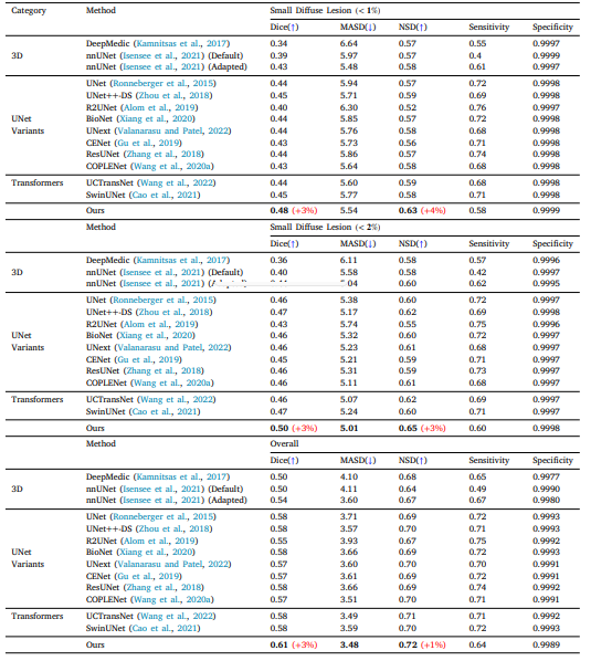
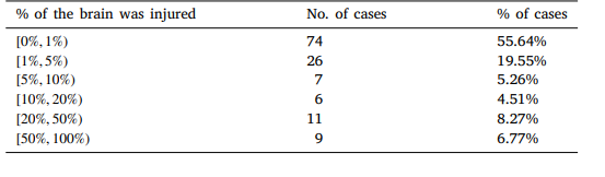
Table 1 HIE dataset. Over 50% of patients with HIE small diffuse lesions.
表 1 HIE 數據集。超過 50% 的 HIE 患者存在微小彌散性病變
Table 2 Performance comparison of ParadiseNet and other deep learning methods for HIE lesion segmentation across a 4-fold cross-testing dataset. ?Results are segmented into overall performance, small diffuse lesions smaller than 1% of the image area, and small diffuse lesions smaller than ?2% of the image area. ParadiseNet shows marked improvements in both Dice, MASD and NSD metrics, particularly in the segmentation of small ?diffuse lesions.
表 2 ParadiseNet 與其他深度學習方法在 HIE 病變分割的 4 折交叉測試數據集上的性能對比。結果分為整體性能、小于圖像面積 1% 的微小彌散性病變和小于 2% 的微小彌散性病變三部分。ParadiseNet 在 Dice、MASD 和 NSD 指標上均表現出顯著改進,尤其在微小彌散性病變的分割中。
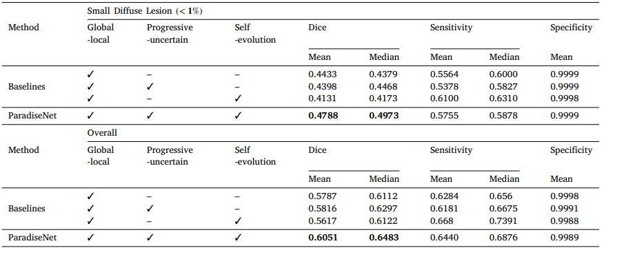
Table 3 A Comparative analysis of segmentation performance between ParadiseNet and baseline models for small diffuse lesion detection in HIE. The ?table presents the Dice coefficient, sensitivity, and specificity metrics for lesions under 1% of the image area, as well as the overall performance ?across the entire dataset. The contributions of global–local learning, progressive uncertainty, and self-evolution modules to the performance ?enhancement in ParadiseNet are also highlighted.
表 3 HIE 微小彌散性病變檢測中 ParadiseNet 與基線模型的分割性能對比。表格展示了圖像面積小于 1% 的病變的 Dice 系數、敏感性和特異性指標,以及整個數據集的整體性能。同時強調了全局 - 局部學習、漸進不確定性和自進化模塊對 ParadiseNet 性能提升的貢獻。
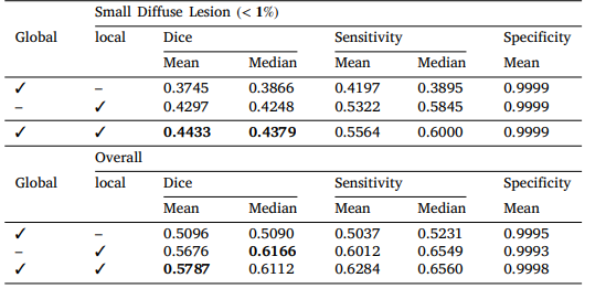
Table 4 HIE 4-fold Ablation Study on global and local stream.
表 4 HIE 全局和局部流的 4 折消融研究
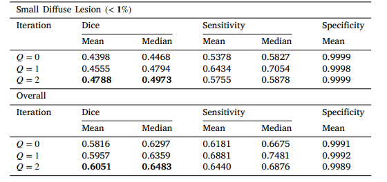
Table 5 HIE 4-fold Ablation Study on Self Iterations.
表 5 HIE 自迭代次數的 4 折消融研究
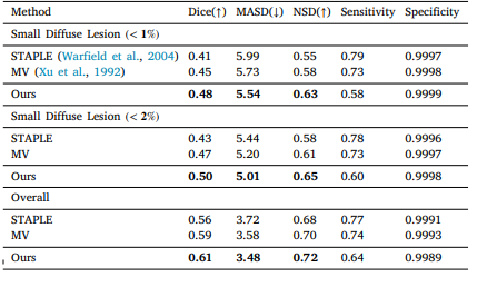
Table 6 Performance comparison of ParadiseNet and ensemble results of top 10 methods in ?Table 2.
表 6 ParadiseNet 與表 2 中前 10 種方法集成結果的性能對比


格式化串口輸出---Prj04)


)

